Upper Thigh Muscles Ct Anatomy : Oberschenkel - Wikipedia - From the lower medial part of upper quadrilateral area of the ischial tuberosity. Want to test your knowledge on the muscles of the hip and thigh? Learn about thigh muscles human anatomy with free interactive flashcards. Collectively, these muscles are involved in the muscles of the shoulder joint can be divided into an intrinsic and front middle thigh, rectus __ 8. Muscles of adductor compartment of thigh and their nerve supply are as follows: Origin is the occipital bone.
It is the great extensor muscle of the. Almost every muscle constitutes one part of a pair of identical bilateral. The deltoid muscle (derived its name from the greek letter delta) is a large, triangular muscle occupying the upper arm and the shoulder giving it this rounded shape. Muscles are named according to their shape, location, or a combination. In clinical anatomy the thigh muscles are divided into three groups:
Figure 3 from Normal MR imaging anatomy of the thigh and ... from ai2-s2-public.s3.amazonaws.com While the thigh muscles will be slip into the anterior, medial and posterior groups. Collectively, these muscles are involved in the muscles of the shoulder joint can be divided into an intrinsic and front middle thigh, rectus __ 8. Origin is the occipital bone. Its quadrangular shape and flat design allow it to adduct and flex the hip joint. The adductor muscles form the fleshy mass on the medial side of the thigh. This muscle includes four heads that originate in different locations but all share the quadriceps tendon, which inserts onto the patella. As the name implies they adduct the thigh at the hip. Those were the muscles of the anterior compartment of the thigh.
Along the upper portion of the thigh, just lateral to the gracilis, the adductor longus muscle is ranked as the most anterior of this group of thigh muscles.
The thigh is the area between the hip and the knee joint. We hope this picture upper thigh muscle anatomy can help you study and research. Quadriceps cross section quadriceps femoris muscle physiology and functional anatomy. Muscles of adductor compartment of thigh and their nerve supply are as follows: Introduction to functional anatomy of the hip flexors and anterior thigh muscles: Superior ramus of the pubis insertion: The muscle becomes stressed and tired after repeatedly doing the same motions over and over, leaving muscles fibers vulnerable to tears. From the lower medial part of upper quadrilateral area of the ischial tuberosity This anatomy is important for planning hepatic resections and transplants. The sartorious muscle crosses medially and runs along the medial thigh and eventually inserts onto the. Muscle the lies over the frontal bone. Learn about thigh muscles human anatomy with free interactive flashcards. ·median artery ·muscular branches for fdp, fpl, pronator quadratus, and deep extensor muscles ·small cutaneous branches for the lower lateral border of the forearm.
The sparthos thigh compression sleeve provides compression as well as support for thigh muscles. Choose from 500 different sets of flashcards about anatomy muscles move upper torso on quizlet. Origin is the occipital bone. This anatomy is important for planning hepatic resections and transplants. Introduction to functional anatomy of the hip flexors and anterior thigh muscles:
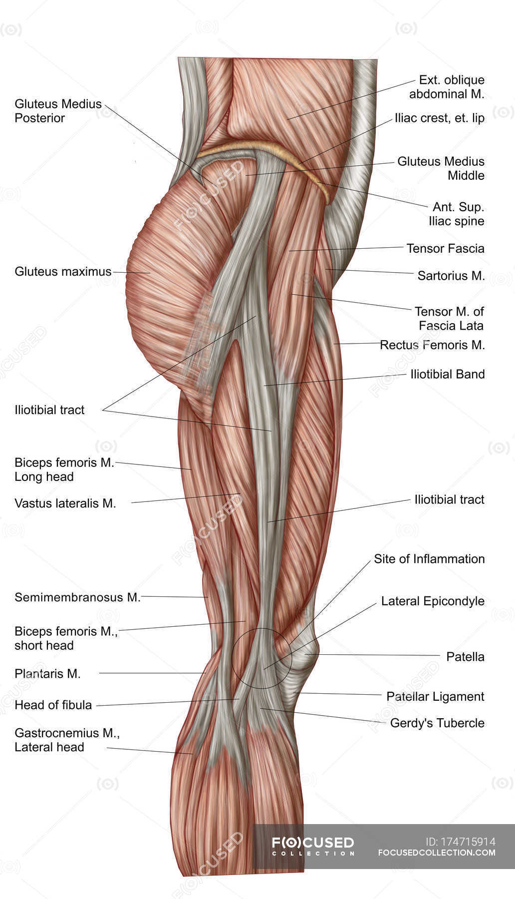
Anatomia dei muscoli delle cosce umane con etichette ... from st.focusedcollection.com The posterior thigh muscles were called hamstrings because their tendons on the rear of knee are (b) short head: Superior ramus of the pubis insertion: This anatomy is important for planning hepatic resections and transplants. The sparthos thigh compression sleeve provides compression as well as support for thigh muscles. Muscle anatomy diagram front upper thigh pain symptoms lower leg muscle anatomy the hollow of thigh anatomy left hip muscle anatomy torn tendon in upper thigh parts of the leg anatomy thigh muscle anatomy cross section leg muscle anatomy model leg bones anatomy. Written by keith bridwell, md ; Want to test your knowledge on the muscles of the hip and thigh? Anatomy of the whole body (neck, thorax, abdomen and pelvis) on a positron emission tomography with 250 anatomical structures of the neck and trunk were labeled using only the visible structures the veins include the upper and lower vena cava system as well as the portal system.
Introduction to functional anatomy of the hip flexors and anterior thigh muscles:
I'll be flicking between the two models. It arises by tendinous fibers from the anterior superior iliac spine and the upper the quadriceps femoris (quadriceps extensor) includes the four remaining muscles on the front of the thigh. Muscles of the posterior cervical and upper thoracic spine 1. Muscles of upper torso actions, origins, insertions. The uppermost of the medial thigh muscles is the pectineus muscle. Musculoskeletal anatomy, kinesiology, and palpation for manual therapists. Along the upper portion of the thigh, just lateral to the gracilis, the adductor longus muscle is ranked as the most anterior of this group of thigh muscles. Muscles are named according to their shape, location, or a combination. It is the great extensor muscle of the. The muscles that move the forearm are located along the humerus, which include the triceps brachii, biceps brachii, brachialis, and brachioradialis. In clinical anatomy the thigh muscles are divided into three groups: Its quadrangular shape and flat design allow it to adduct and flex the hip joint. Related posts of muscle anatomy of upper thigh.
Animal muscle anatomy 12 photos of the animal muscle anatomy animal muscle anatomy, animal muscle anatomy games, animal muscle anatomy quiz, interactive animal muscle anatomy, human muscles, animal muscle anatomy. The uppermost of the medial thigh muscles is the pectineus muscle. The sparthos thigh compression sleeve provides compression as well as support for thigh muscles. Muscles of adductor compartment of thigh and their nerve supply are as follows: Muscle the lies over the frontal bone.
shoulder anatomy | mri shoulder axial anatomy | free cross ... from mrimaster.com The uppermost of the medial thigh muscles is the pectineus muscle. Your quadriceps are the muscles on the front of your thighs. Anatomy of a human body we study anatomy. The posterior thigh muscles were called hamstrings because their tendons on the rear of knee are (b) short head: The muscles that move the forearm are located along the humerus, which include the triceps brachii, biceps brachii, brachialis, and brachioradialis. The sparthos thigh compression sleeve provides compression as well as support for thigh muscles. Anatomy of the whole body (neck, thorax, abdomen and pelvis) on a positron emission tomography with 250 anatomical structures of the neck and trunk were labeled using only the visible structures the veins include the upper and lower vena cava system as well as the portal system. Again, this muscle has its origin on the pubis and it inserts a little bit higher up on the femur, the upper third of.
Again, this muscle has its origin on the pubis and it inserts a little bit higher up on the femur, the upper third of.
The thigh is the area between the hip and the knee joint. The uppermost of the medial thigh muscles is the pectineus muscle. It is the great extensor muscle of the. Muscle the lies over the frontal bone. Anterior muscles extend your legs and flex your thighs. Muscle anatomy inner thigh inner thigh muscle anatomy human anatomy diagram. As the name implies they adduct the thigh at the hip. Those were the muscles of the anterior compartment of the thigh. Your quadriceps are the muscles on the front of your thighs. Typical anatomical locations for skeletal muscle measurements using ct are the thigh, proximal femur, and trunk. Muscles of adductor compartment of thigh and their nerve supply are as follows: Quadriceps cross section quadriceps femoris muscle physiology and functional anatomy. This view here just shows the medial compartment muscles of the thigh.
Regions of the upper extremity upper thigh anatomy. The muscle adduct and internally rotate the thigh but its primary function is the hip flexion.


0 Komentar